Kidney cancer is the ninth most commonly occurring cancer in men and the 14th most commonly occurring cancer in women. Renal cell carcinoma (RCC), also known as renal cell cancer or renal cell adenocarcinoma, is the most common type of kidney cancer (about 9 out of 10 kidney cancers). They arise from structures called Renal Tubules which filter fluids in the kidney before the waste (urine) is discharged into the bladder. About 5 to 10 percent of kidney cancers are Transitional cell cancers (TCC). These don’t start in the kidney itself, but in the lining of the renal pelvis (where the ureters meet the kidneys). These are usually more aggressive than RCCs. Most people diagnosed with kidney cancer are between 65 and 74. Kidney cancer is twice as common in men than in women, and African Americans, American Indian and Alaskan Natives are more likely to be diagnosed.
What are the risk factors for RCC?
The following risk factors have been associated with RCCs:
How is RCC diagnosed?
Many early kidney cancers are discovered during testing for other reasons. A urine sample may show blood, or a blood test may detect anemia. An imaging study of the abdomen for unrelated symptoms may reveal kidney cancer.
If doctors suspect kidney cancer, imaging studies are important for diagnosis. In some cases, kidney cancer may be diagnosed with imaging studies alone.
These include:
If surgery is recommended, the diagnosis also may be confirmed post-op by looking at cancer cells under a microscope, called pathologic diagnosis. Kidney cancer also may be confirmed with a needle biopsy.
Learn more about diagnosing kidney cancer
What are the treatment options available?
Surgery is the first-line treatment option for most patients with renal cell carcinoma. Other treatment options for all types of kidney cancer include:
Prostate cancer is the most common cancer and the second leading cause of cancer death among men in the United States. The American Cancer Society (ACS) estimates that about 248,530 new cases of prostate cancer will be diagnosed in 2021. Prostate cancer usually grows very slowly, and finding and treating it before symptoms occur may not improve men’s health or help them live longer.
Some prostate cancers are aggressive. Approximately one in every 41 men diagnosed will die from the disease, according to the ACS. Black men are reportedly more prone to developing fast-growing prostate cancers that start causing problems earlier and are harder to treat.
What are the risk factors of prostate cancer?
Prostate cancer is rarely diagnosed in men younger than 40. By age 50, men undergo changes in the size and shape of the cells in the prostate. Understanding whether these changes are signs of a tumor and knowing your risk for developing prostate cancer are important steps in protecting your health.
Besides age, other risk factors for prostate cancer include:
What are the common signs and symptoms of prostate cancer?
In the early stages, prostate cancer usually doesn’t show symptoms. However, as prostate cancer grows, it may lead to:
Patients who develop new or concerning symptoms should consult with their doctor or urologist.
Learn more about the signs and symptoms of prostate cancer
How is Prostate Cancer diagnosed?
Symptoms are often absent in the early stages of prostate cancer. Many cases are discovered through routine screening tests. Getting screened for prostate cancer is an individual decision. It may help to discuss the risks and benefits with a doctor.
Screening for prostate cancer usually involves the following tests:
If either of these suggests the possibility of prostate cancer, doctors typically perform additional tests before making a diagnosis.
The only way to know for sure whether a tumor is cancerous is by examining cells under a microscope, a procedure also known as a prostate biopsy.
Learn about diagnostic procedures for prostate cancer
What are the treatment options?
Deciding on prostate cancer treatment is a personal decision made between a patient and his care team. Factors such as preferences, age, health history and the cancer stage all play a role in the decision-making process.
Treatment may involve one or a combination of these options:
Other therapies that are used less commonly, or are not considered standard treatment for prostate cancer, include:
Learn more about treatments for prostate cancer
Testicular cancer most often begins in germ cells (cells that make sperm). It is uncommon and is most frequently diagnosed in men ages 20 to 34. Most testicular cancers can be cured, even if diagnosed at an advanced stage.
What are the risk factors?
These include:
Of the factors associated with the risk of developing germ cell tumours of the testis, cryptorchism and malignancy in the contralateral testis are by far the strongest. Testicular microlithiasis, vasectomy, and scrotal trauma are not risk factors for testicular cancer.
What are the common symptoms of testicular cancer?
The common symptoms of testicular tumor are:
How is testicular cancer diagnosed?
Patients should seek an urgent consultation if they suspect abnormalities in the testis. The common diagnostic tests are:

What are the treatment options?
Management is dependent on the type of tumour and stage. Approximately 90% of patients classified as having a good prognosis achieve a durable complete remission with treatment. Even metastatic disease should be seen as potentially curable. When treating young adults with a highly curable disease, possible long-term toxicity of treatment is an important consideration. Following confirmation of a germ cell tumour, all patients should be referred to a specialist centre for the management of testicular tumours.
Where possible, a radical orchidectomy should be performed. A testicular prosthesis should be offered to all patients. When appropriate, sperm storage should be offered to men who may require chemotherapy or radiotherapy.
Patients with metastases where the diagnosis is not in doubt (high tumour markers and the presence of a testicular mass) should be referred for immediate chemotherapy
Do patients need follow-up after treatment?
All patients post treatment of testis cancer will need a strict follow-up. Your doctor will schedule you for regular visits after treatment to see if the cancer has come back (recurrence). Visits may include a physical examination, blood tests, chest x-ray, and/or a CT scan to look for new tumors. Visits are usually more frequent just after treatment ends. Follow-up typically continues for at least 5 years. If there is no recurrence, you do not need further treatment.
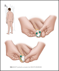
Examining your testicles is easy
Examine your testicles regularly (Fig. below), especially if you have a risk factor for testicular cancer.
The best time to examine your testicles is right after a hot bath or shower. The scrotal skin will be relaxed, and the testicles can be felt more easily. It takes only a few minutes.
Do the exam standing:
If you find a lump, swelling, or other change, see you doctor. Changes are not always cancer, but if it is cancer and you catch it early, you have the best chance of cure.
Bladder cancer is a common type of cancer that involves the bladder. Most often the cancer starts in the cells (urothelial cells) that line the inside of your bladder. Urothelial cells are also found in the kidneys and the tubes (ureters) that connect the kidneys to the bladder. Urothelial cancer can also happen in the kidneys and ureters, although less frequent than in the bladder.
What cause bladder cancer?
Till date it is unclear what causes bladder cancer. A number of risk factors have been identified, but in many cases none of them may be present. (Risk factor is not a cause in itself but increases the risk that a cancer may occur). The risk factors are:
How is bladder cancer diagnosed?
Tests and procedures used to diagnose bladder cancer may include:
After confirming bladder cancer, additional tests may be recommended to determine whether the cancer has spread to your lymph nodes or to other areas of your body. These include:
What are the available treatment options?
Treatment options for bladder cancer depend on a number of factors, including the type of cancer, grade of the cancer and stage of the cancer, overall health of the affected individual and his treatment preferences
The options are:
Surgery, to remove the cancer cells. Common types of surgery for bladder cancer are:
Surgery, to remove the cancer cells. Common types of surgery for bladder cancer are:
Transurethral resection of bladder tumor (TURBT). An endoscopic surgery to diagnose and remove cancers confined to the inner layers of the bladder, usually performed for cancers that aren’t yet muscle-invasive cancers. This may be performed using laser technology.
Cystectomy. A surgery to remove all or part of the bladder that may be undertaken by open, laparoscopic or robotic technology. Partial cystectomy is a procedure where the surgeon removes only the portion of the bladder that contains a single cancerous tumor. Radical cystectomy is an operation to remove the entire bladder and the surrounding lymph nodes. After radical cystectomy a new way of urine storage and expulsion needs to be created using segments of intestine, called urinary diversion (different techniques are neobladder, ileal conduit, continent diversion).
A combination of treatment approaches may be recommended by your doctor and members of your care team.
What measures can be followed to reduce the risk of bladder cancer?
Although there’s no guaranteed way to prevent bladder cancer, few steps can help to reduce bladder cancer risk.
 +973 66907199